OQ PathScope
Light Engine, Scanner, and Integrated PC
Optional: Workstation Package: Scanner Stand, Monitor, Keyboard & Mouse
High resolution OCT for ex-vivo tissue. It’s OCT. Re-imagined
Optical Coherence Tomography, better known as OCT, uses light to provide cross-sectional images of micro-layers of tissues and other samples. OCT is a valuable bench tool for Pathologists who want to work smarter and more efficiently with their histological sections.
It’s improving pathology
Allows for multi-scale lateral resolution and field of view, which helps you more thoroughly explore your ex-vivo samples.
Saves time by helping you select fewer, more targeted histological sections.
It’s affordable
Costs a fraction of any other OCT system, allowing many pathology labs to now afford this technology.
It’s compact
About the size of a shoe box, so it won’t crowd your laboratory bench — or require a special cart.
It’s accurate
Delivers amazing depth and transverse resolution, allowing pathologists to generate precise image data.
It’s easy to use
Focus on science, not on setup. The software simple, so you can generate images quickly.
It’s compatible
Integrates with ours or any C-mount-equipped microscope (custom C-mount configuration at no additional cost)..
Technical Data
| Image Size | 512 px x 512 px |
| Depth Resolution | 4,5 μm in air 3,0 µm in tissue |
| Transverse Resolution | 4x: 10 μm; 10x: 5 μm; 40x: 2 µm |
| Scan Range | 4x: 5 mm; 10x: 2,5 mm; 40x: 1 mm |
| A-Scan Line Rate | 8,800/sec |
| B-Scan Image Rate | 12/sec |
| Center Wavelength | 880 nm |
| Sensitivity (OSNR) | 100 dB |
| Output Power | 750 μW |
| System Size (cm) | 33,0 x 19,1 x 15,2 |
| Scanner Size (cm) | 11,4 x 6,4 x 4,8 |
| System Weight | 2,72 kg |
Samples OCT Images

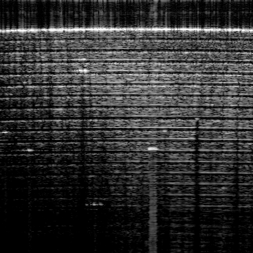
Layers of Scotch Tape
Alternating layers of tape and adhesive captured by OQ PathScope.

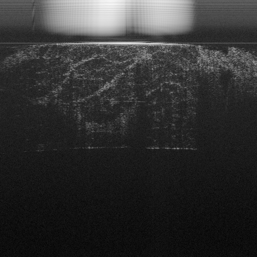
Bacon
The OQ PathScope allows you to distinguish between fat tissue on the left and muscle tissue on the right.

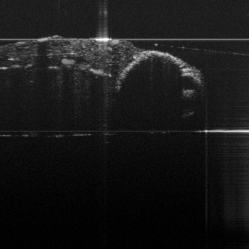
Zebra Fish
Seen from above using OQ PathScope You can see its protruding eyes on the right.
