Crystal arthritis
The introduction of computed tomography (CT) has contributed to major advances in medical imaging. CT imaging has taken plain x-ray radiographs from two-dimensional flat images to three-dimensional images. Three-dimensional imaging provides information about the structure more clearly and illustrates the relationship between adjoining structures. Color CT has the potential to be as revolutionary to medical imaging as the move from two-dimensional images to three-dimensional images. MARS offer new and improved diagnostic information at significantly lower radiation dose compared to conventional, black and white CT systems. This will significantly broaden the value and use of CT as both a research tool and in clinical diagnostic imaging.
L. Stamp et al. “Clinical utility of multi‐energy spectral photon‐counting CT in crystal arthritis.” Arthritis Rheumatol, 71: 1158-1162. doi:10.1002/art.40848 (2019)
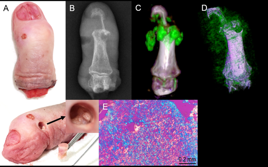
MARS Microlab 5X120 provides multi-energy information and crystal type specific images of small joints with increased diagnostic value at higher spatial resolution (<80 µm) compared to traditional modalities.
A. An excised gouty finger with a core (arrow) revealing subcutaneous Tophus.
B. Plain x-ray showing bone erosions with overhanging edges and soft tissue changes consistent with tophus.
C. Conventional dual-energy CT image and D. high resolution MARS scanner, both depicting monosodium urate (MSU) crystal deposits, with MARS detecting finer detail and higher MSU volume.
E. Polarized light microscopy of a sample obtained from the core, confirming the presence of negatively birefringent MSU crystals.
Atherosclerotic plaque
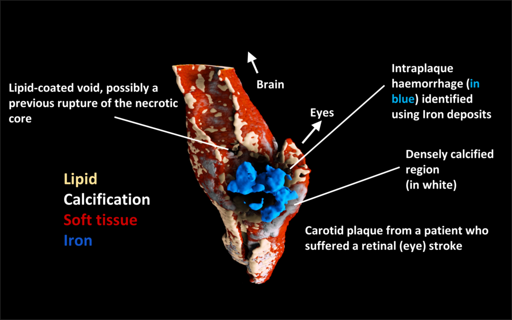
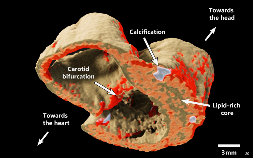
Plaque specimens showing intra plaque haemorrhage in blue along densely calcified regions. The image on the right is an exposure rendered image of an excised human carotid atheroma at the bifurcation level showing lipid-like, calcium-like, and water-like material components. The darker the color hue, the greater the quantity of the component.
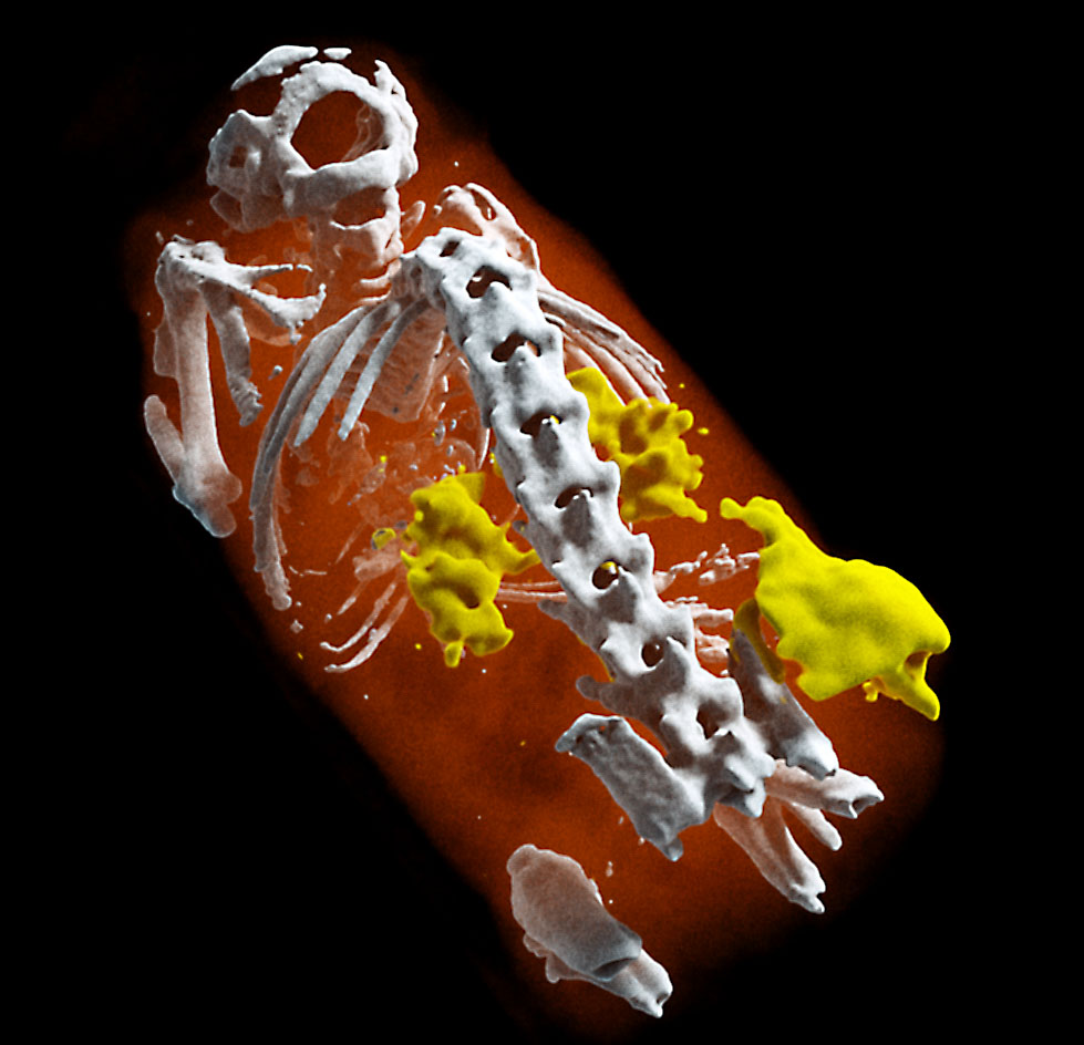
Functionalized nanoparticle contrast
Another promising application of spectral x-ray imaging is the use of functionalized nanoparticle contrast. The functionalization of nanoparticles refers to the addition of a molecule so that the nanoparticle can perform a specific action. For example, tumor targeting with an antibody molecule. Efficient targeting of diseased cells, while healthy cells remain untreated, will make a substantial contribution towards precision medicine. MARS Microlab 5X120 scanners offer groups developing pharmaceuticals an opportunity to non invasively image and measure normal and pathological processes, drug delivery, and response to treatment.
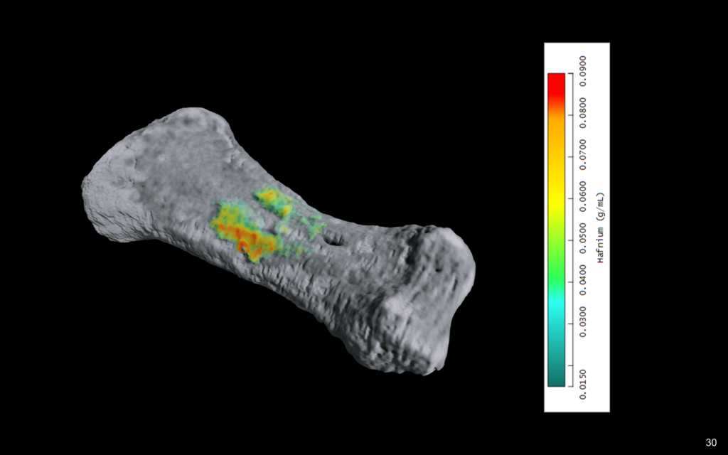
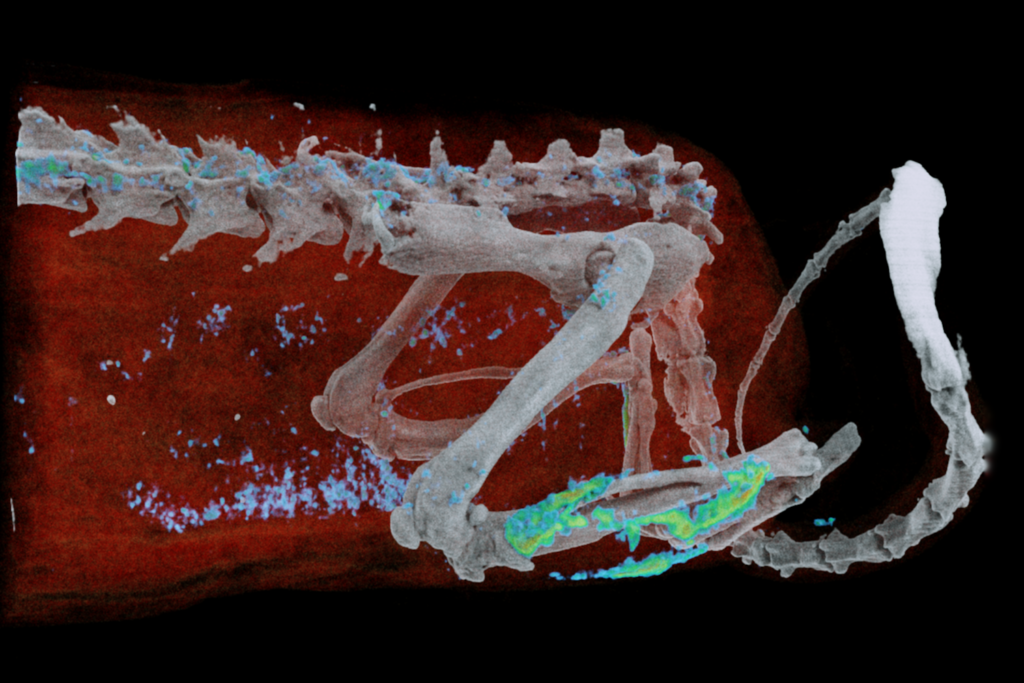
Identification and quantification of a novel hafnia-based functionalized nanoparticle. Hafnia was materially separated from bone (calcium) and soft tissues (water and lipid). This nanoparticle was used to highlight bone microdamage in an excised human finger bone (left) and rat specimens (right). Further, functionalized nanoparticles were evaluated versus non-functionalized nanoparticles. This study was in collaboration of Mars Bioimaging with the University of Maryland, Baltimore, USA and the University of Illinois at Urbana–Champaign, Urbana, USA.
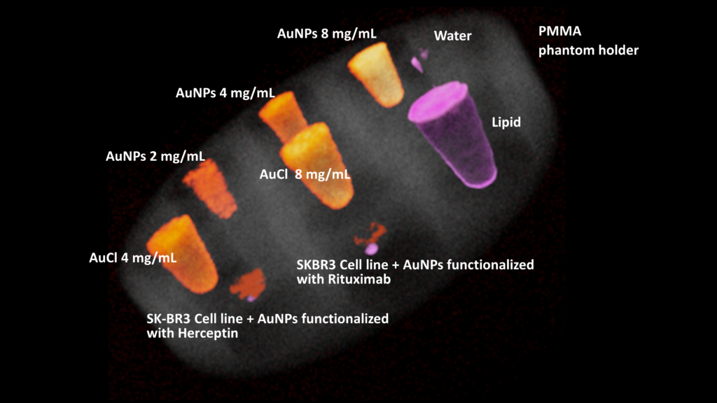
Using MARS Microlab 5X120, preclinical methodology to quantify drug delivery to specific cell types was established. MARS reconstruction and material decomposition algorithms can identify and quantify low concentrations of gold nanoparticles labeled with a monoclonal antibody drug.
M. Moghiseh et al. “Spectral photon-counting molecular imaging for quantification of monoclonal antibody-conjugated gold nanoparticles targeted to lymphoma and breast cancer: an in vitro study.” Contrast media & molecular imaging. (2018)
Increased attenuation in lung lesions
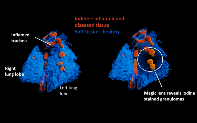
With high-resolution and high-soft tissue contrast of MARS material images, the local increased attenuation (due to increased density of parenchyma) in the lung may be identified and quantified earlier and more accurately than conventional CT images. This improves the detectability of ground-glass opacities, crazy paving appearances and air space consolidation which are important radiographic features in the diagnosis of tuberculosis and pneumonia.
Similarly, with high-resolution MARS attenuation images, peribronchovascular interstitial thickness, bronchial wall thickness and lumen size of bronchi and bronchioles may be measured more accurately than conventional CT images, which ultimately improve the detection of TB earlier.
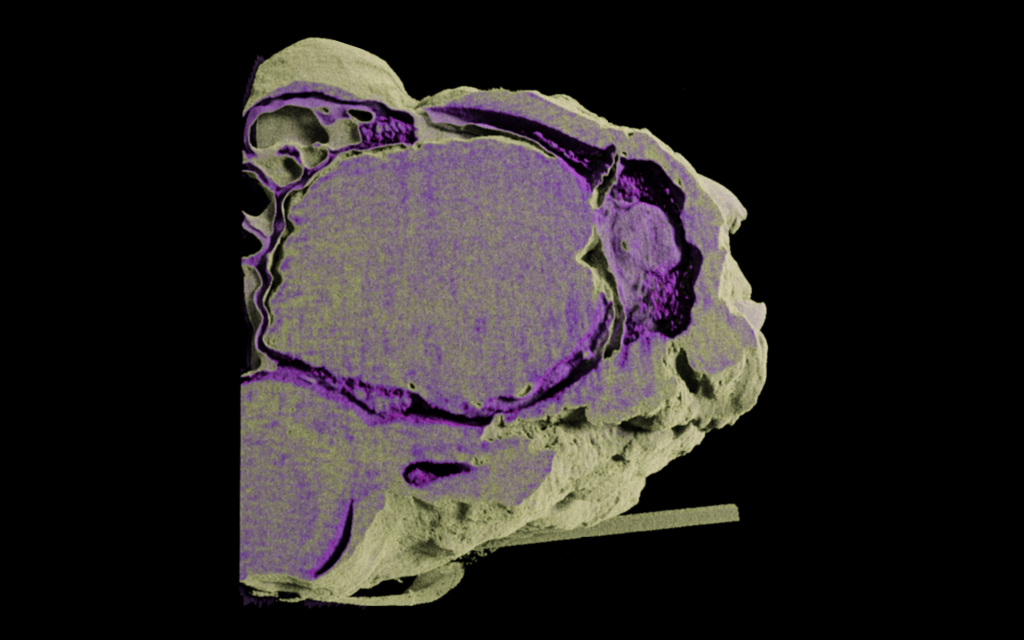
Brain structure changes
The structure of white matter (WM) and grey matter (GM) can change in response to different brain diseases. Improving WM and GM differentiation, along with monitoring anatomical changes at high spatial resolution can lead to an increase in the specificity and sensitivity of the detection of brain diseases. In the case of imaging a brain stimulator electrode, the metal artifact produced by the electrode largely distorts image quality in conventional CT and MRI images. Thus, the benefits of spectral photon-counting CT include improved soft tissue differentiation and reduced metal artifact. Additionally, high resolution provides a platform for investigating the minute bone features within the head.
Left: sheep brain with implanted electrode. Right: sheep brain showing WM and GM differentiation.
Tumor angiogenesis
Use of high-Z materials as imaging contrast agents could provide the exact location of disease, measure drug delivery, and monitor the response to treatment, all in a single scan.
This image shows 15 nm non-functionalized gold nanoparticles used as a contrast agent to distinguish angiogenesis. If the gold nanoparticles had a drug attached, then drug delivery could be targeted to tumor angiogenesis.
Detection of arthroplasty implant failure
Summary by lead Author, Dr Lawrence Lau:
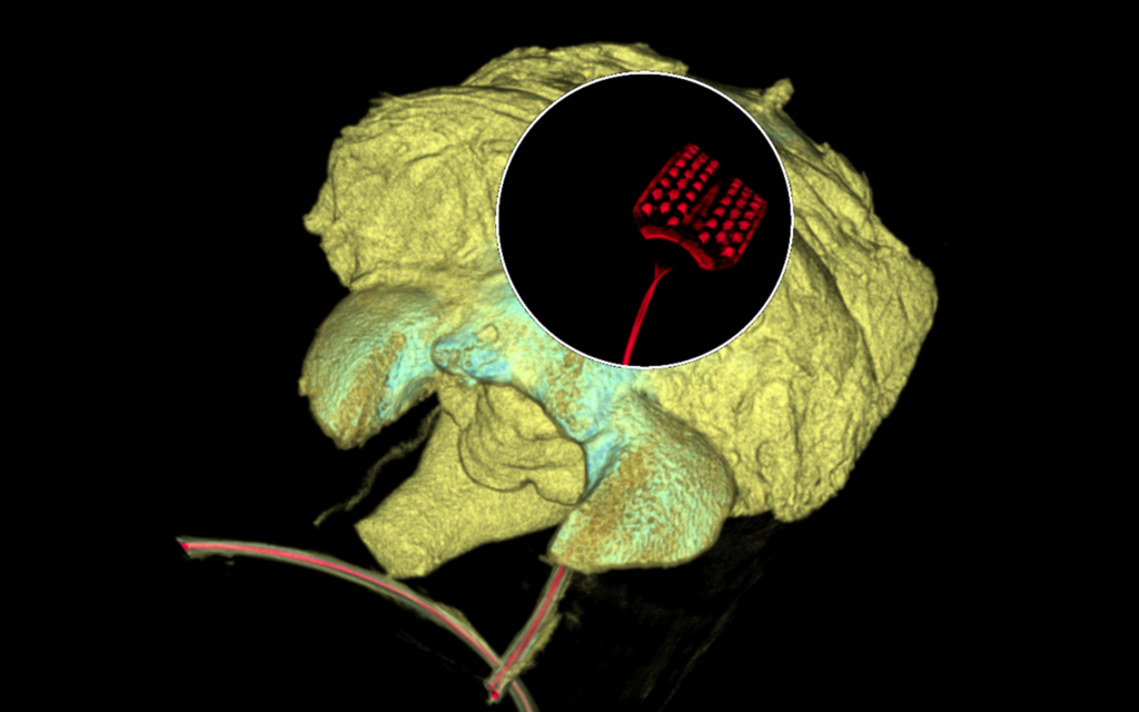
In the recent article; “Multi-energy spectral photon-counting computed tomography (MARS) for detection of arthroplasty implant failure” by Lau LCM, Lee WYW et. al., in Scientific Reports, our team published the first report on using the technology of MARS CT in the field of orthopedics. Through MARS CT, we were able to demonstrate the polyethylene and tibial tray wear of a failed total knee arthroplasty (TKA), while pre-revision radiographs, ultrasound, and MRI of the same TKA could not identify such wear. We shall continue to work on this investigatory track to better characterize the use of MARS CT in detecting orthopedics implant failure and integration with human bodies, and hopefully will integrate this technology in our clinical practice very soon.
Lau, L.C.M, et al., (2021). Multi-energy spectral photon-counting computed tomography (MARS) for detection of arthroplasty implant failure. Scientific Reports, vol. 11, 1554. https://doi.org/10.1038/s41598-020-80463-2: Read paper
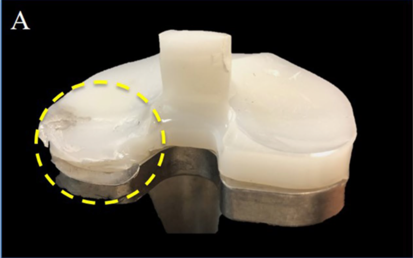
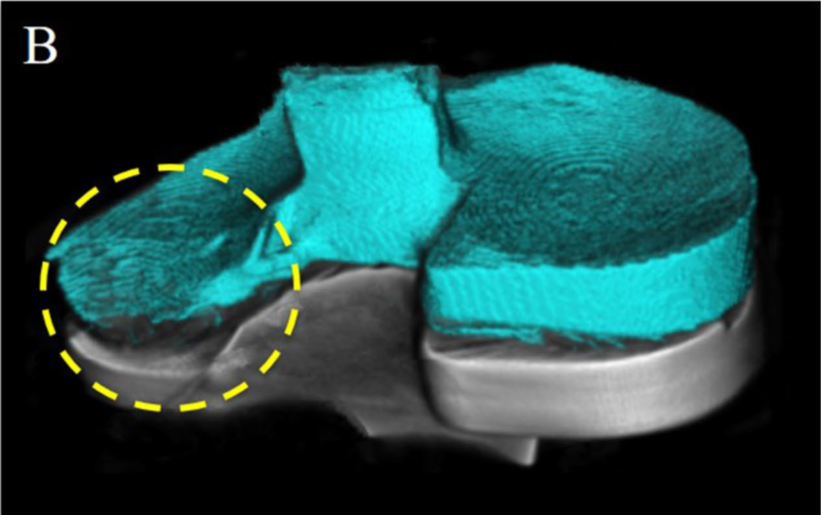
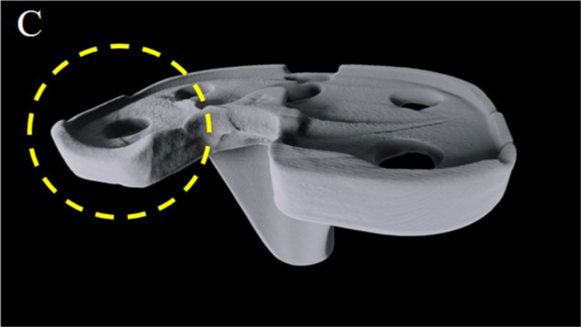
(A) Clinical photos, (B) MARS-CT images and (C) MARS-CT images with polyethylene digitally removed. The wear at the postmedial aspect of the polyethylene implant and tibial metallic tray is clearly visible (circle).